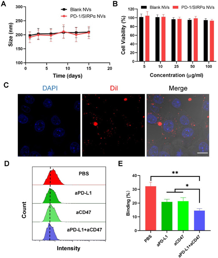
FIGURE 2.
In vitro bioactivity analysis of PD-1/SIRPα NVs. (A) Stability analysis of PD-1/SIRPα NVs in PBS solution after different days of incubation. The nanovesicles were incubated in PBS with constant shaking at 100 rpm at 37°C. (B) Biocompatibility analysis of hybrid nanovesicles on DC2.4 cells by measuring the cell viability of treated cells. DC2.4 cells were treated with Blank NVs or PD-1/SIRPα NVs for 48 h at 37°C. The cell viability of treated cells was analyzed via CCK8 assay. (C) Cellular uptake of PD-1/SIRPα NVs in melanoma cells. B16F10 cells were incubated with Dil-labeled PD-1/SIRPα NVs for 2 hours at 37°C. Scale bar: 10 μm. (D, E) Flow cytometry curves and quantitative analysis of the blockade property of PD-1/SIRPα NVs. Melanoma cells were pretreated with PBS, anti-PD-L1 antibodies (aPD-L1), anti-CD47 antibodies (aCD47), or a combination of aPD-L1 and aCD47 for 4 hours at 37°C. After washing with PBS, Dil-labeled PD-1/SIRPα NVs were added to each well for a 2-hour incubation. Treated cells were digested and subjected to flow cytometry analysis (gated on PE).

