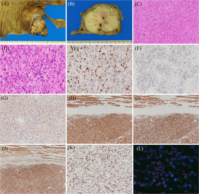
Figure 3.
Histopathological findings of dedifferentiated liposarcoma showing a leiomyosarcoma phenotype. Resected specimens demonstrate that the tumor with lobulated and gray-white color (A, B) has spindle cells with eosinophilic cytoplasm and mitotic figures (C, D). Immunohistochemical results for the indicated proteins are shown: (E) MDM2, (F) CDK4, (G) S-100, (H) h-caldesmon, (I) desmin, (J) α-SMA, and (K) Ki67. Fluorescence in situ hybridization shows MDM2 amplification (L).

