Fig. 1.
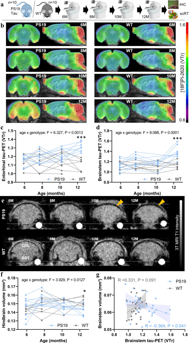
Monitoring of tau pathology and atrophy in PS19 and wild-type mice using [18F]PI-2620 PET/MRI. a Experimental workflow of serial PET/MRI imaging sessions and terminal immunohistochemistry (IHC) and single-cell Radiotracing (scRT) in PS19 and wild-type (WT) mice. b Coronal and axial group average [18F]PI-2620 PET images obtained using an MRI template show the monitoring of radiotracer binding (volume of distribution ratios, VT; striatal reference), with pronounced temporal and brainstem patterns in aged PS19 mice (n = 8–10) compared with WT mice (n = 8–10). c, d Mixed linear models of entorhinal and brainstem [18F]PI-2620 PET signals indicate a significant age × genotype effect and elevated tau PET signals in PS19 mice compared with WT mice at 12 months of age. e Examples of serial MRI atrophy patterns in an individual PS19 mouse compared with a WT mouse. Coronal slices are shown with indications of the cerebellum (CBL) and brainstem (BST) regions. The orange arrows highlight atrophy in PS19 mice. f Mixed linear model of hindbrain volume indicating a significant age × genotype effect and a decreased hindbrain volume in PS19 mice compared with WT mice at 12 months of age. g Association between tau PET signals and brain volume in the brainstem of PS19 and WT mice across all investigated time points showing greater atrophy in the presence of high tau PET signals, specifically in PS19 mice

