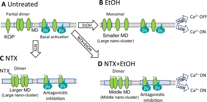
Fig. 5. Model of NTX and ethanol effects on KOP lateral organization and interactions with lipids.
A In untreated cells, KOP largely diffuses freely in the lipid bilayer, with a small fraction that localizes in cholesterol-enriched membrane domains (MDs) and forms homodimers. KOPs that localize in MD can be activated by thermal fluctuations, even without the presence of an externally introduced agonist. Through this basal activation of KOP, KOP-mediated inhibition of Ca2+ channel can take place. B Under treatment with EtOH, deformation of MD is taking place, KOP dimers dissociate, forming monomers that freely diffuse in the lipid bilayer. This, in turn leads to reduction in the KOP-mediated inhibition of the Ca2+ channel, and the Ca2+ channel is in the ON-state under lower EtOH concentrations (0 mM–80 mM). Under high EtOH concentration (>100 mM), plasma membrane deformation is excessive and the Ca2+ channel may be generally inhibited. C Under the treatment with NTX, larger nanoscale KOP clusters form (based on evidence from [8]). NTX-bound KOP forms predominantly homodimers in larger MDs. The Ca2+ channel is released from the inhibiting action of KOP by NTX binding. D EtOH-modulated lipid dynamics is suppressed by NTX. Middle-sized nano-clusters are being formed. NTX also exerts its antagonist activity at KOP, thus the Ca2+ channel is released from KOP-activated-inhibition.

