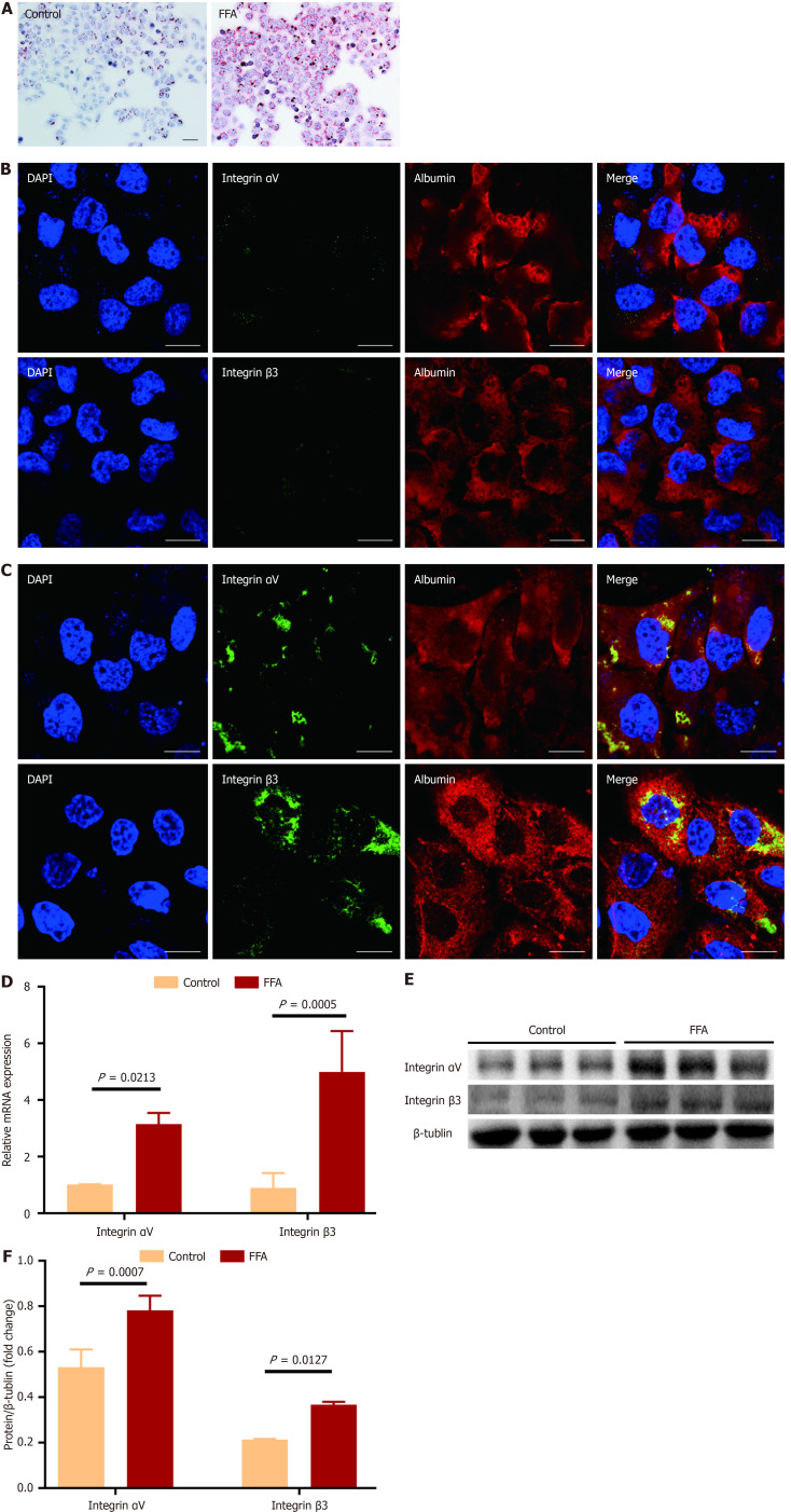
Figure 1.
Expression of integrin αvβ3 in cultured hepatocytes. Human LO2 hepatocytes were cultured with the medium containing 100 μmol/L palmitate acid and 200 μmol/L oleic acid (FFA) for 24 hours, and the cells cultured in the medium without FFA served as the control. A: Representative micrographs of the control and FFA-cultured hepatocytes after stained with Oil-Red O. Images were taken at original magnification (200 ×), scale bars = 20 μm; B and C: Representative fluorescent images of the control (B) and FFA-cultured (C) hepatocytes after separately stained with integrin αv and β3 subunits antibody (green color) and counterstained with albumin antibody (red color). 6-diamidino-2-phenylindole was used for nuclei staining. The merged images show the yellow color area by overlaying images of the counterstaining. Images were taken at original magnification (400 ×), scale bars = 100 μm; D: Comparison of the message RNA levels of integrin αv and β3 subunits in the control and FFA-cultured hepatocytes. The message RNA levels of integrin αv and β3 subunit were determined by quantitative real-time polymerase chain reaction analysis; E and F: Comparison of the protein amounts of integrin αv and β3 subunits in the control and FFA-cultured hepatocytes. The protein amounts of integrin αv and β3 subunits were analyzed by western-blot assay, and β-Tublin was used as the reference. All experiments were undertaken in triplicates. In all panels, data are expressed in means ± SD. FFA: Oleic acid; DAPI: 6-diamidino-2-phenylindole.

