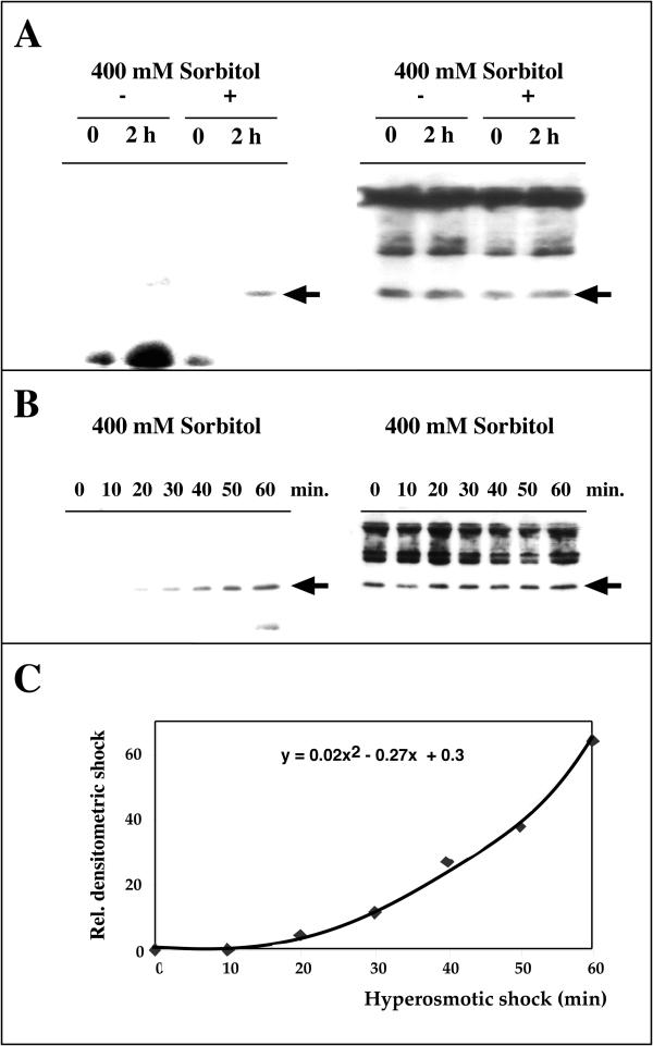
Figure 2.
Hisactophilin is phosphorylated in wild type cells exposed to hyperosmotic conditions (A). Cells were metabolically labeled with 32P-orthophosphate prior to hypertonic shock. Hisactophilin was immunoprecipitated from samples withdrawn at T = 0 and T = 2 h. The probe was separated by SDS-PAGE and blotted. Autoradiography (left panels A,B) was performed for 48 h and hisactophilin was subsequently identified by immunostaining (right panels A, B). (+) denotes samples from shocked cells, (-) denotes samples from control cells suspended in SPB buffer. The arrow indicates the position of 15 kDa proteins. (B) The amount of phospho-hisactophilin progressively increased in hyperosmotically shocked cells. The experiment was performed as described in (A). (C) Densitometric analysis of the autoradiography in (B) reveals an exponential increase in phospho-hisactophilin upon hyperosmotic stress: the kinetics can be fitted by a quadratic function displayed in the figure. The densities are given in relative arbitrary units, setting the background as 0.

