. 2024 Nov 8;12:RP90695. doi: 10.7554/eLife.90695

Figure 4. Human-induced pluripotent stem cell (iPSC)-derived microglia demonstrate robust phagocytosis.

Human iPSC-derived microglia were incubated for 1 hr in pHrodo Red E. coli bioparticles (A), pHrodo Red zymosan bioparticles (B), DiI-labeled bovine photoreceptor outer segments (POSs) (C) and labeled with anti-human P2RY12 antibody (green) and DAPI. Scale bar = 40 µm. (D) A high-magnification view of a POS-containing intracellular vesicle within a labeled microglial cell is shown. Scale bar = 40 µm. (E) An overlay of panels in (D) with side views. Scale bar = 40µm.

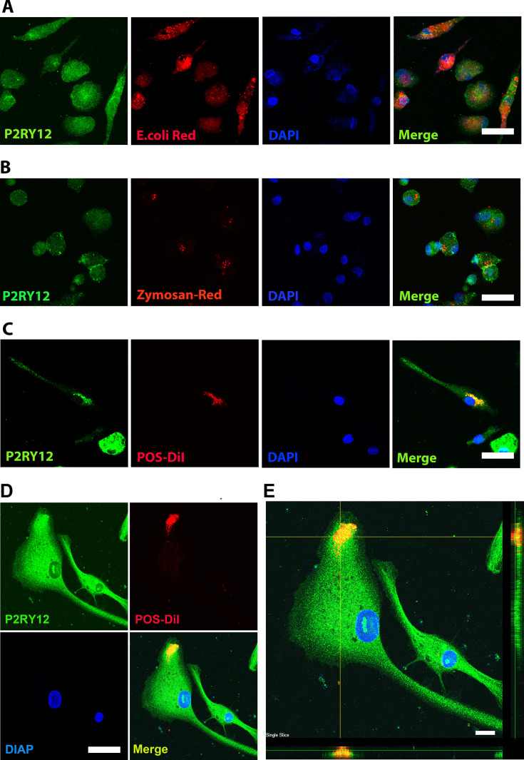
Figure 4.

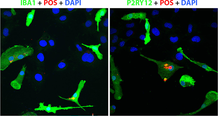
Figure 4—figure supplement 1. Floating myeloid progenitor cells were cultured in a 2-well slide chamber overnight, and then the DiI-labeled bovine photoreceptor outer segments were added to the chamber and incubated for 1 hr.

Figure 4—figure supplement 1.

The cells were fixed with 4% paraformaldehyde (PFA) for 20 min and stained with IBA1 and hP2RY12, respectively. The DiI-labeled bovine photoreceptor outer segments can only be engulfed by IBA1- or P2RY12-positive microglia cells. Scale bar = 16 µm.
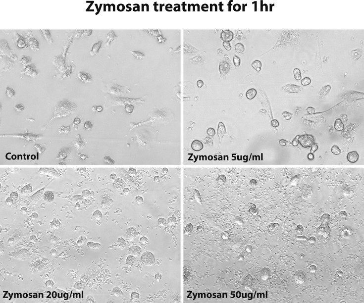
Figure 4—figure supplement 2. The morphology of human-induced pluripotent stem cell (hiPSC)-derived microglia cells under 1 hr zymosan treatment with different concentrations.

Figure 4—figure supplement 2.

After 1 hr of treatment with zymosan, 5 µg/ml concentration did not change the microglia cell morphology, but over 20 µg/ml concentration changed the microglia morphology to an ameboid round shape.