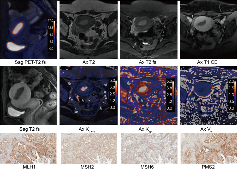
Fig. 5.
The PET/DCE-MRI images and immunohistochemistry of a 48-year-old female with MMRd endometrioid carcinoma (FIGO IA stage, grade 1). An intrauterine mass showed significant glucose hypermetabolism (SUVmax = 28.73, SUVmean = 11.40, MTV = 13.71, TLG = 156.39) and hyper blood flow perfusion (Ktrans = 4.2, Kep = 11.06, Ve = 0.37). a Sagittal PET and T2 fs fused image. b Axial T2-weighted image. c Axial T2 fs image. d Axial T1 CE image. e Sagittal T1 CE image. f Axial Ktrans map. g Axial Kep map. h Axial Ve map. i MLH1 protein immunohistochemical staining (× 100). j MSH2 protein immunohistochemical staining (× 100). k MSH6 protein immunohistochemical staining (× 100). l PMS2 protein immunohistochemical staining (× 100). T2 fs T2-weighted fat suppression, T1 CE T1-weighted contrast enhanced, SUVmax maximum standardized uptake value, SUVmean mean standardized uptake value, MTV metabolic tumor volume, TLG total lesion glycolysis, Ktrans transfer constant, Kep efflux rate, Ve extravascular extracellular volume

