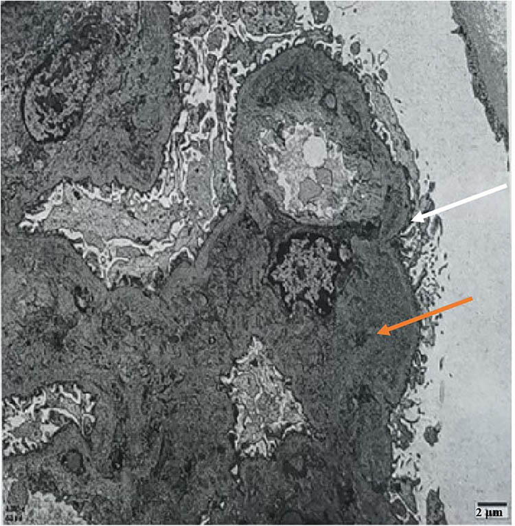
Figure 4.

Electron microscope, white arrows indicate segmental mesangial interposition, where mesangial cells or matrix appear to insert into the capillary loop, altering the normal glomerular architecture. Orange arrows point to blocky and clustered electron-dense material in the mesangial area and subendothelial segments, which could represent immune complex deposits or other pathologic accumulations.
