Fig. 2.
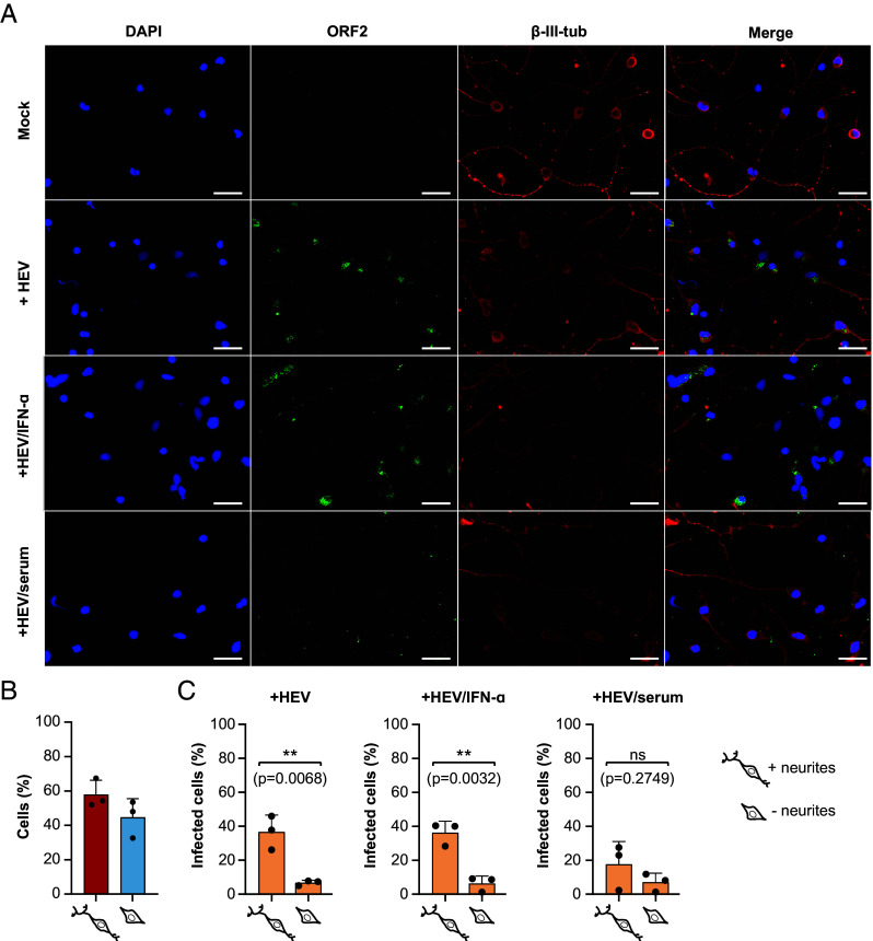
Infection of human-iPNs with HEVcc Kernow-C1 p6 strain. (A) Immunofluorescence staining was performed for uninfected (Mock) and infected iPNs without treatment or with IFN-α (1,000 Units/mL) or human anti-HEV serum. DAPI was used to stain the nuclei (blue), a polyclonal rabbit anti-HEV antiserum to stain the ORF2-encoded capsid protein (green) and an anti-β-III-tubulin (β-III-Tub) antibody to stain the neuronal cytoskeleton. (Scale bars represent 50 µm.) (B) The percentage of neurons with or without neurites were determined by quantifying the β-III-tubulin signal. (C) The susceptibility of neurons, both with and without neurites, to HEV infection was quantified by measuring the ORF2 signal using CellProfiler. An unpaired t test was used to assess statistical significance with a significance level of α = 0.05. ns: not significant.

