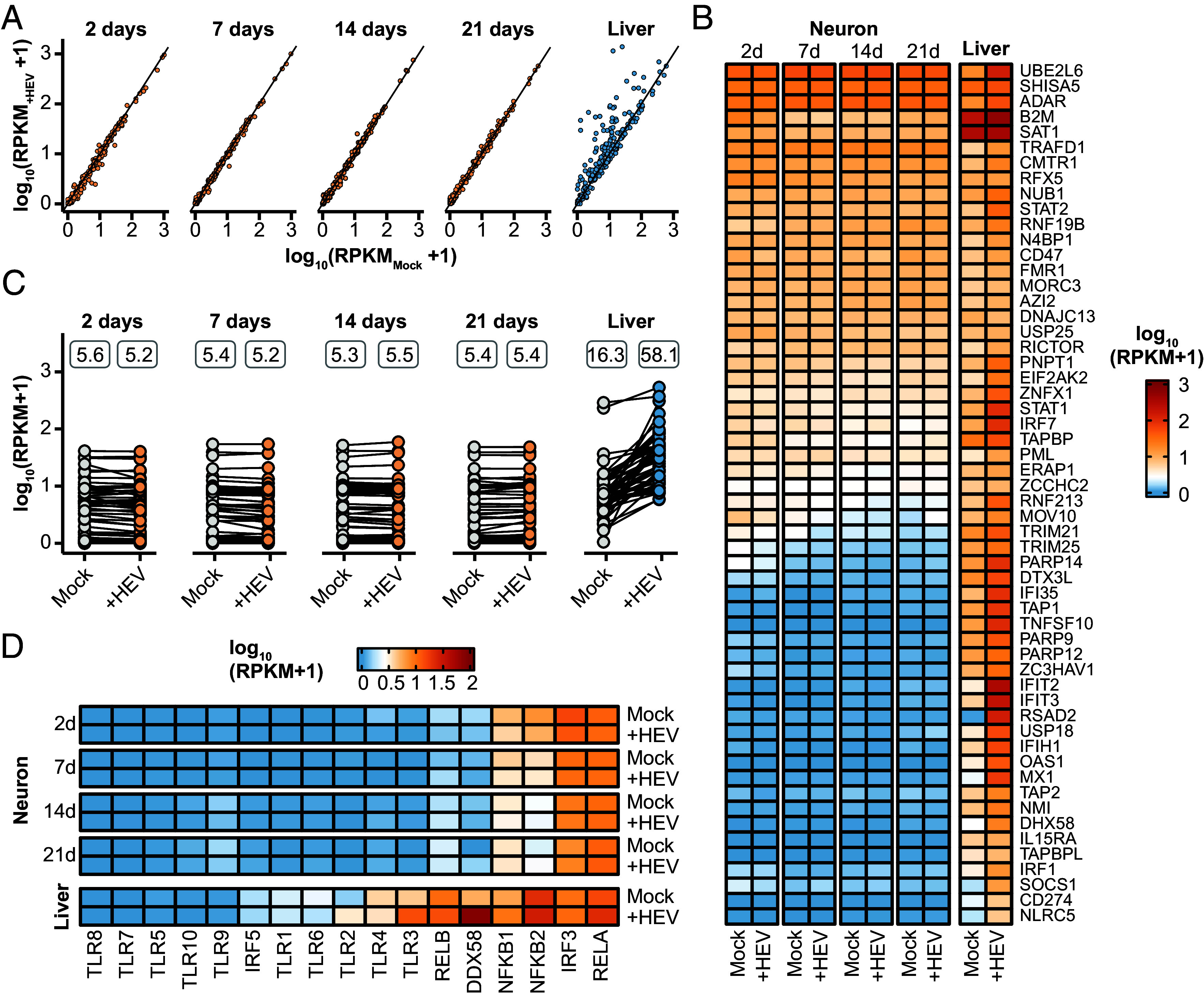
Fig. 4.

Neuronal intrinsic expression of ISGs and absence of innate immune response to HEV infection. (A) Overview of the general immune response. Depicted are all genes within the GO term immune response (GO:0006955) of uninfected (x-axis) and infected (y-axis) neurons at different differentiation time points (2, 7, 14, 21 d) and primary human hepatocytes (PHH, liver). The primary neurons (orange) were infected with HEV for 5 d and the PHHs (blue) were infected for 48 h. (B) Expression pattern of core ISGs of uninfected (Mock) and infected (+HEV) primary neurons at different differentiation stages and PHH samples. Color-code represents log10 RPKM+1 values. (C) Overview and comparative analysis of the expression levels of core ISGs measured in RPKM values, in both uninfected (Mock) and HEV-infected (+HEV) primary neurons and PHH. The values above displayed represent the mean RPKM expression levels of all genes under each respective condition. (D) Expression pattern of pathogen-sensing genes including Toll-like receptors of Mock and HEV-infected iPNs and PHH.
