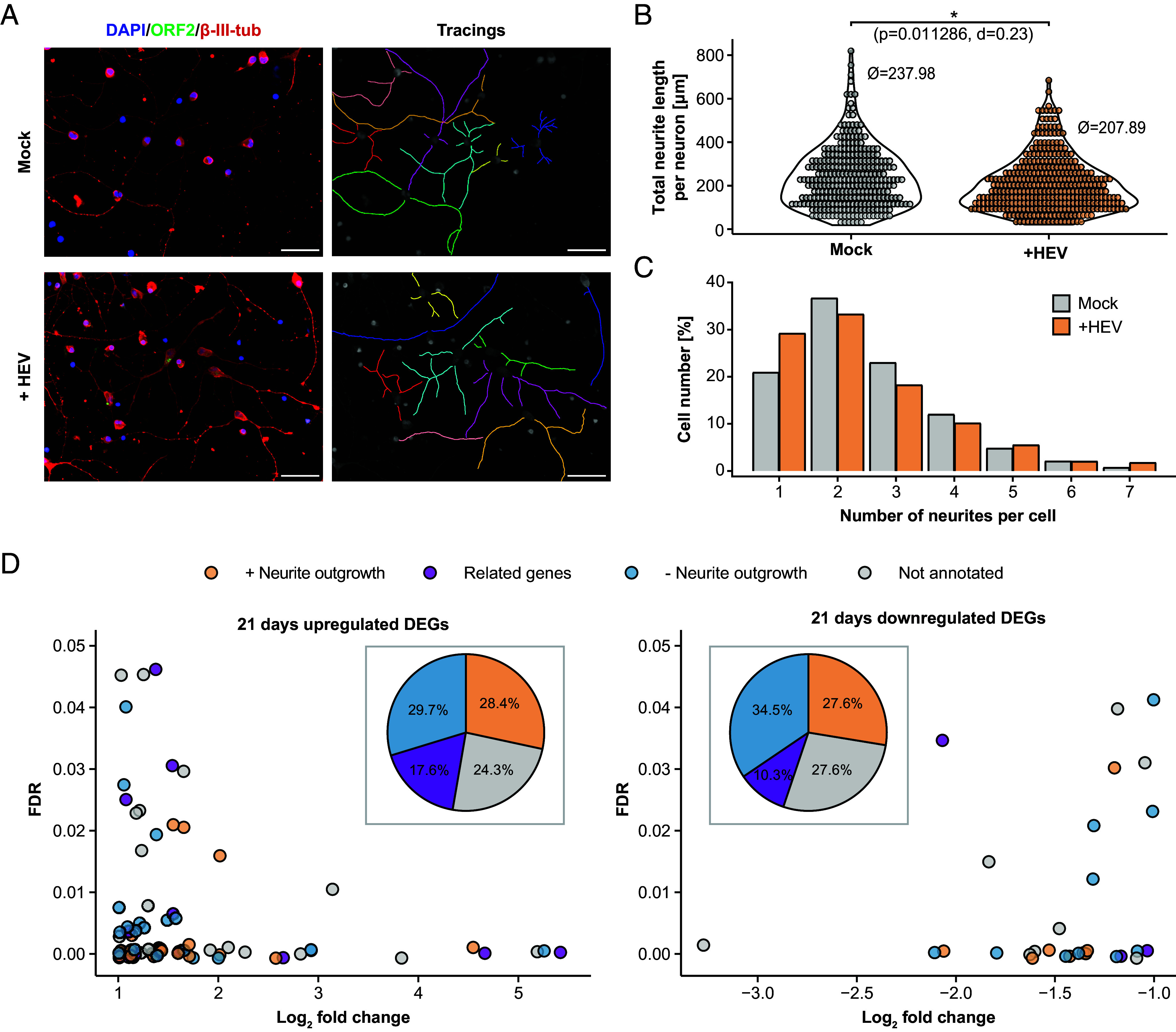
Fig. 5.

Reduction of neurite length of iPNs after HEV-inoculation. The effect of HEV inoculation on iPNs neurite length compared to mock-inoculated cells (21-d-old cells) was examined through microscopic analysis. Five days postinoculation, cells were stained with DAPI (blue, nucleus) and with antibodies against the ORF2 capsid protein (green) and β-III-tubulin (red, cytoskeleton). ImageJ’s NeuronJ plugin was used to manually track the neurites of each neuron (A) and determine the total length of neurites per neuron (B) and the number of neurites per cell (C). To test the significance of differences in neurite length, Mann–Whitney U test followed by Bonferroni P-value correction was used. The effect size was calculated as Cohen´s d (d). (Scale bars represent 100 µm.) (D) A literature review was conducted on the 72 up- and 29 down-regulated genes identified in 21-d-old HEV-infected cells to determine whether these genes were previously reported in connection with neurite outgrowth (orange), had related genes (family members) associated with neurite outgrowth (purple), had no known relation to neurite outgrowth, or were not annotated (SI Appendix, Tables S1 and S2). The pie chart represents the percentage of these categories relative to the total number of significantly up- and down-regulated genes.
