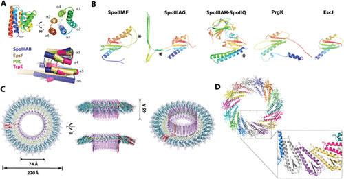
FIGURE 2.

Structures of the core components of the sporulation channel share similar structural motifs with homologs from other bacterial secretion systems. (A) (Top) The SpoIIIAB soluble domain adopts a six-helix bundle fold with both N and C termini in close proximity and facing the mother cell membrane. The molecule is shown in two views, related by a 90° rotation. (Bottom) SpoIIIAB shares a fold similar to that of homologous proteins from the T2SS and T4PS. Shown is a structural overlay of SpoIIIAB with EpsF, TcpE (both from Vibrio cholerae), and PilC (Thermus thermophilus) proteins in blue, wheat, green, and pink, respectively (PDB codes 6BS9, 3C1Q, 2WHN, and 4HHX, respectively). Two regions of structural variation are seen in the helix 6 angle and the increasing dimensions of helices 4 and 5 and the loop connecting them. (B) SpoIIIAF, SpoIIIAG, and the SpoIIIAH-SpoIIQ heterodimer contain an RBM fold similar to that of the T3SS basal body proteins, EscJ (Escherichia coli) and PrgK (Salmonella Tryphimurium) (PDB codes 6DCS, 5WC3, 3UZ0, 1YJ7, and 3J6D, respectively). All five structures are displayed in cartoon representation and rainbow color scheme and for clarity are individually shown in identical orientations originating from structural superposition. SpoIIIAF is presented as an overlay of the two monomers seen in the crystal structure, with the region of alternate conformation associated with regulation marked with an asterisk. SpoIIIAG adopts the canonical RBM fold, with a large insertion of the β-triangle motif marked with an asterisk. An SpoIIIAH additional N-terminal helix is marked with an asterisk. (C) Cryo-EM structure of the SpoIIIAG soluble domain 30-meric ring. A three-dimensional reconstruction and atomic model are shown in top side, cropped, and tilted views. The SpoIIIAG ring structure is colored according to distinctive ring elements: RBM in cyan, planar β-ring in green, and vertical β-ring in pink, with the single protomer in red. (D) SpoIIIAH-SpoIIQ representative computational modeled ring, here in C15 symmetry with zoomed-in view of the predicted interaction region between the RBMs of SpoIIIAH. Ring model coordinates were obtained from Meisner et al. (46).
