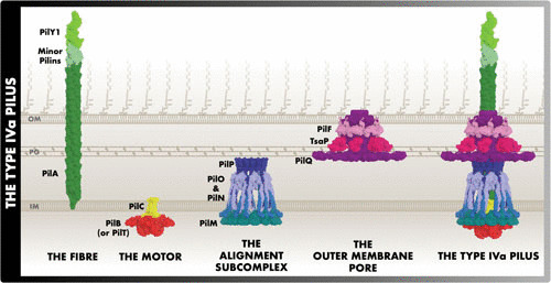
FIGURE 1.

Subcomplexes of the T4aP. The protein structures portrayed reflect the full-length structure predictions and their predicted location in the T4aP. This figure is largely consistent with the previously published working model of the M. xanthus T4aP (43). Due to limited information, there is uncertainty regarding the locations of PilF, TsaP, PilY1, and the minor pilins.
