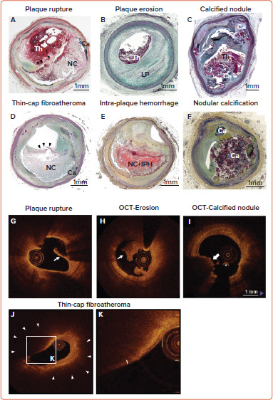
Figure 1: Pathology and Intra-coronary Imaging (OCT) of Human Coronary Artery Morphologies Associated With ACS.

Pathologic (A–F) and OCT-based classification (G–K) of human coronary artery associated with ACS. A: plaque rupture; B: plaque erosion with underlying pathologic intimal thickening; C: calcified nodule; D: thin-cap fibroatheroma (black arrowhead shows thin fibrous cap); E: intra-plaque hemorrhage; F: nodular calcification; G: OCT-plaque rupture (white arrow shows disrupted thin fibrous cap); H; OCT-erosion (white arrow shows white thrombus); I: OCT-calcified nodule (white arrow shows overlying superficial calcification with red thrombus); J–K: thin-cap fibroatheroma. J shows low-power image. K is high-power magnification of the rectangular area in J. The white arrow head in J shows low backscattering, signal-poor region with diffuse border, suggesting large necrotic core. The double-head white arrow in K shows thin fibrous cap.
ACS = acute coronary syndrome; Ca = calcification; IPH = intra-plaque hemorrhage; LP = lipid pool; NC = necrotic core; OCT = optical coherence tomography; Th = thrombus. Source: Panels G, H, J and K: Otsuka et al. 2014.[12] Adapted with permission from Springer Nature. Panel I: Jia et al. 2013.[13] Adapted with permission from Elsevier.
