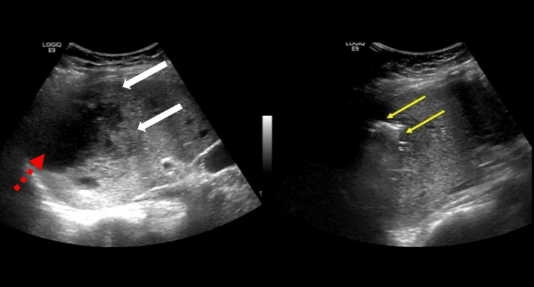
Figure 2. Ultrasound scan of the abdomen showing a large irregular hypoechogenic cavity (red broken arrow) with a solid component at the boundary (white arrows) located in the dome of the diaphragm. A different section of the lesion showed irregular echogenic shadows with mild posterior reverberation artifacts (yellow arrows) of air collections consistent with a GFPLA.
GFPLA: gas-forming pyogenic liver abscess

