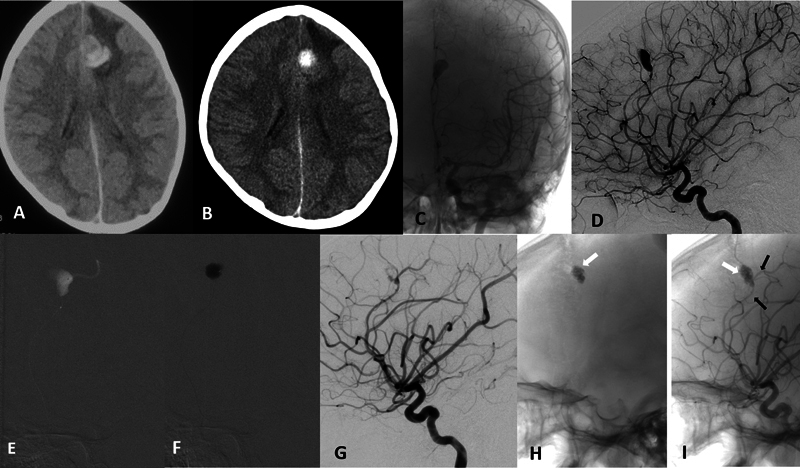
Fig. 4.

Case-3. ( A ) Initial axial noncontrast computed tomography (NCCT) of the brain showing intracerebral hemorrhage (ICH) in the left frontal lobe in a para-alpine location without any mass effect. ( B ) Repeat NCCT done at the time of admission showed minimal resolution of hematoma. ( C ) Native anteroposterior (AP) and ( D ) subtracted digital subtraction angiography (DSA) images of the left internal carotid artery (ICA) injection show a dissecting aneurysm of the left distal pericallosal artery. ( E ) Fluoroscopic image of microcatheter injection in the left pericallosal artery proximal to the aneurysm showing the aneurysm and distal artery. ( F ) Fluoroscopic image showing the glue injected within the aneurysm sac with sparing of the neck to preserve the parent artery. ( G ) Immediate postglue DSA run showing near complete obliteration of aneurysm with minimal staining of the wall. Delayed native DSA image ( H ) without contrast and ( I ) after contrast showing the glue cast ( white arrow ) with complete aneurysm occlusion and preserved parent artery ( black arrows ).
