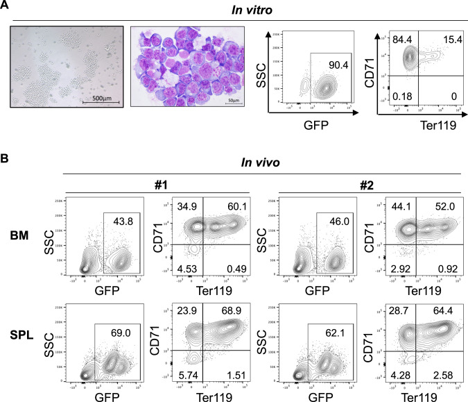
Fig. 4. Establishment of a murine AEL cell line: CEP53.
A CEP53 cells in suspension (far left), Wright-Giemsa staining of a cytospin slide of CEP53 cells (left) and flow cytometric analysis of CEP53 cells for GFP (right) and Ter119 and CD71 (far right). B CEP53 cells were transplanted into non-irradiated recipient mice. Bone marrow (BM) and spleen (SPL) cells were collected from two moribund mice. Expression of GFP, Ter119, CD71 in BM and SPL cells were shown. Note that almost all GFP+ CEP53 cells were CD71+Ter119+/− erythroblasts.

