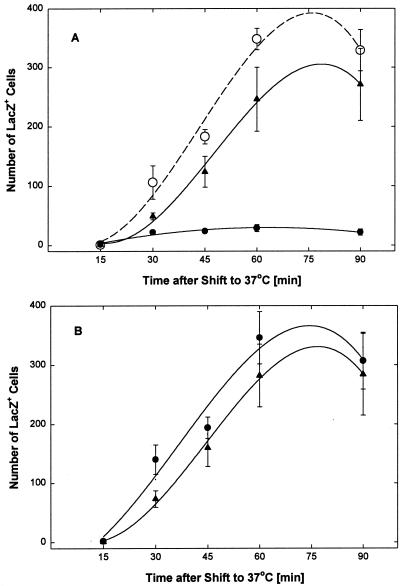
FIG. 8.
EB blocks virus entry. Virus (2.6 × 10³ PFU of hrR3 per well) was preadsorbed for 1 h at 4°C to Vero cells (2 × 10⁵ cells/well in 96-well microtiter strip plates), and the cultures were switched to ice-cold serum-free DMEM and kept for an additional 1 h at 4°C before they were transferred to 37°C to initiate virus entry. Every 15 min following the temperature switch, strips of wells were treated for 1 min with low-pH citrate buffer to inactivate any remaining extracellular virus. After each citrate treatment, cultures were returned to serum-supplemented medium. (A) EB (●) or EBX (▴) at 25 μM was added immediately after preadsorption of the virus and remained present until the citrate treatment. Mock-treated controls were kept in peptide-free medium (○). (B) EB (●) or EBX (▴) at 50 μM was added immediately after each citrate treatment and remained present until all cultures were fixed and stained for β-galactosidase 8 h after the temperature shift to 37°C. Blue cells in areas of 6.5 mm2 were counted in triplicate wells. All data points are means with standard errors of the means.

