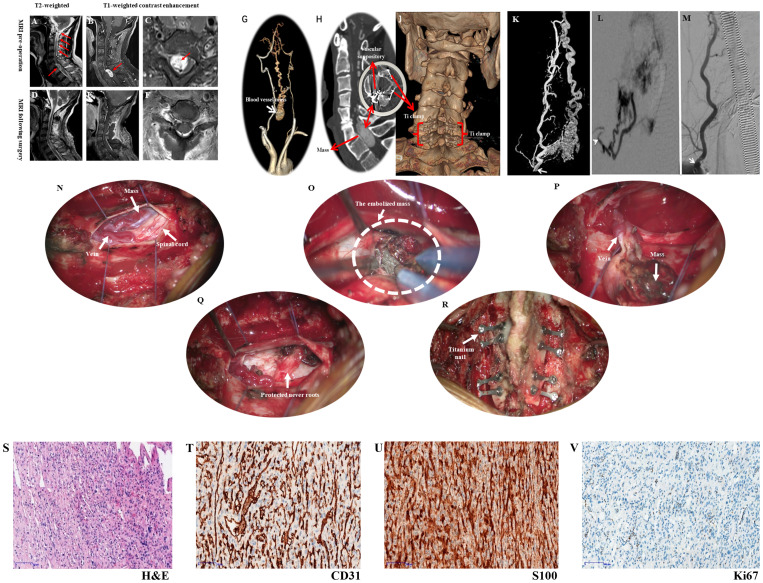
Figure 1.
Presents the imaging examination, surgical procedure, and pathological findings. Figure 1 (A) MRI shows a mass at the C6 and C7 levels, along with a beaded vascular flow shadow on the dorsal side of the spinal cord above it; (B, C) MRI reveals oval abnormal signal foci in the spinal canal at the level of C6-7 vertebral bodies, measuring approximately 16mm×11mm×26mm, exhibiting evident enhancement, as well as small patchy vascular flow shadows (red arrows) and changes indicative of spinal cord compression; (D–F) Depict images from postoperative period illustrating complete disappearance of the mass when compared to (A–C); (G) The CTA reveals a vascular mass located between the bilateral vertebral arteries, potentially supplied by the superior thyroid artery; (H) Preoperative CT scan exhibits a rounded mass with lightly increased signal intensity within the spinal canal at C6-7; (I) Postoperative CT demonstrates complete disappearance of the mass, revealing residual shadows of the embolic agent; (J) Three-dimensional CT reconstruction confirms satisfactory fixation of vertebral plates in laminoplasty using titanium nails. (K) Selective angiography involved injecting a contrast agent through the distal end of the 4F VER catheter, entering the opening of the right thyroid neck trunk (white arrow). This depicted contrast agent entry into the tumor via the intervertebral foramen, causing hyperstaining and backflow towards the medulla oblongata; (L) The Echelon10 microcatheter was super selected into a branch of the tumor feeding artery from the superior thyroid artery (white arrow), resulting in tumor staining after contrast injection; (M) Following Onyx18 injection, angiography of the right thyrocervical trunk displayed well-developed main trunk arteries (white arrow) with nearly complete absence of tumor staining. (N) The dissection of the dura allows for the observation of varicose veins, tumors, and compression of the spinal cord; (O) After cutting the arachnoid membrane and flowing out the cerebrospinal fluid, the embolized tumor can be seen on the lateral side of the spinal cord; (P) The tumor has been dissociated and turned out of the tumor cavity; (Q) The nerve root is completely preserved; (R). Reposition and titanium needle fixation of the lamina fix the vertebral plate, and maintain the stability of the spine. Pathological findings (×100 magnification) observed under a light microscope, (S) H&E staining revealed numerous fusiform tumor cells with irregular lumens and thin-walled blood vessels; (T) CD31 staining showed prominent vascular endothelial cells, with the presence of numerous irregular thin-walled blood vessels and some anastomosed blood vessels; (U) S100 expression was strongly positive in tumor cells; (V) Ki67 expression indicated a low tumor growth index of approximately 1%.

