FIGURE 2.

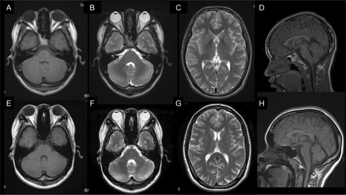
Brain MRI findings of Patient 1 and Patient 2. Brain MRI revealed normal results pertaining to the cerebellar (A–C, E–G) and cerebral hemispheres (D, H). (A–D): Patient 1. (E–H): Patient 2. MRI, magnetic resonance imaging.

Brain MRI findings of Patient 1 and Patient 2. Brain MRI revealed normal results pertaining to the cerebellar (A–C, E–G) and cerebral hemispheres (D, H). (A–D): Patient 1. (E–H): Patient 2. MRI, magnetic resonance imaging.