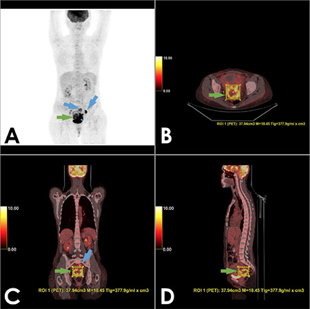
Figure 2.

Maximum intensity projection (MIP) PET (A), fused transaxial (B), coronal (C) and sagittal (D) PET/CT images of a 33 year-old woman with squamous cell uterine cervix carcinoma. She had a primary tumor with SUVmax: 18.45, MTV: 37.94 cm3 and TLG: 377.9 g/mL x cm3 with pelvic lymph node metastases. After PET/CT imaging, she underwent TAH + BSO following by adjuvant chemo-radiation therapy. She had recurrent disease at the 7th month and died at the 12th month after PET/CT
PET: Positron emission tomography, CT: Computed tomography, SUVmax: Maximum standardized uptake value, MTV: Metabolic tumor volume, TLG: Total lesion glycolysis, TAH + BSO: Total abdominal hysterectomy with bilateral salpingooophorectomy, ROI: Region of interest
