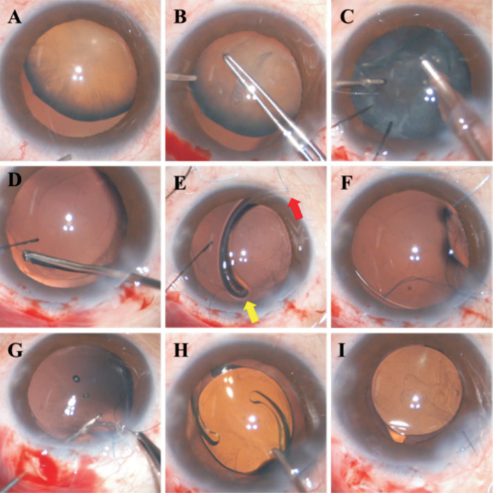
Figure 1. Intraoperative view of the surgical procedures.
A: A subluxated lens with a zonular abnormality of about 180 degrees; B: Continuous curvilinear capsulorhexis was performed gently; C: Phacoemulsification and cortex aspiration were performed with the aid of capsular hooks; D: A 23-gauge vitrector was introduced into the capsular bag through the capsulorhexis to create a 1.5-2 mm diameter capsulotomy at the capsular equator toward the middle location of the zonular abnormality; E: Attached a double-strand 8-0 polypropylene suture (red arrow) at the middle point of a standard CTR and temporarily threaded another guiding suture (yellow arrow) through the leading eyelet of the CTR; F: Delivered the CTR into the capsular bag and aligned the middle point of it to the equatorial capsulotomy made earlier; G: Introduced the needle of the 8-0 polypropylene suture into the hollow needle through the capsulorhexis and passed it through the scleral pocket bed with the guidance of the hollow needle; H: A foldable IOL was implanted into the capsular bag; I: Well-positioned IOL at the end of the surgery. CTR: Capsular tension ring; IOL: Intraocular lens.

