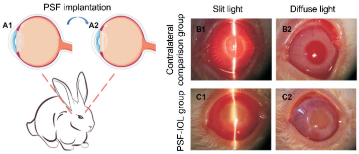
Figure 3. PSF implantation into the rabbit eye and slit-lamp images of contralateral comparison (n=9) and PSF-IOL groups (n=9) under slit and corresponding diffuse light.
A1-A2: Schematic illustration of the PSF implantation. B1-B2: Slit lamp photographs of the contralateral comparison group. B1 is the contralateral comparison group under slit light; B2 is the contralateral comparison group under diffuse light. C1-C2: Slit lamp photographs of the PSF-IOL group. C1 is the PSF-IOL group under slit light; C2 is the PSF-IOL group under diffuse light; PSF: Poly(dimethyl siloxane)-SiO2 thin films; IOL: Intraocular lens.

