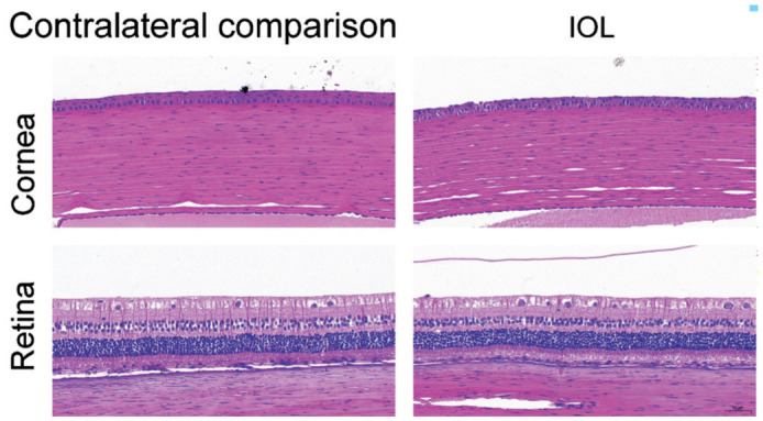
Figure 6. The HE staining of New Zealand white rabbit eyes demonstrating detailed histological identification of the cornea and retina in the contralateral comparison (n=9) and PSF-IOL groups (n=9).

PSF: Poly (dimethyl siloxane)-SiO2 thin films; IOL: Intraocular lens.
