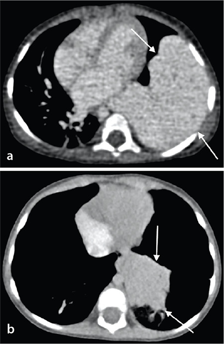
Figure 3.

(a, b) A nine-month-old girl with hemangioma. (a) The axial contrast-enhanced computed tomography (CT) image shows large, homogenous contrast-enhanced tumor in the left lower lobe (arrows). (b) Following one year of propranolol treatment, a contrast-enhanced chest CT showed a significant decrease in the overall tumor size (arrows).
