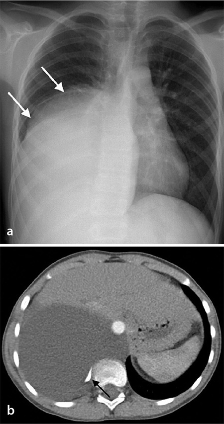
Figure 5.

(a, b) An 11-month-old boy with mature cystic teratoma. (a) The chest X-ray shows large opacity in the lower zone of the right lung. (b) The axial chest computed tomography image demonstrates a large cystic mass with calcification (black arrow) and a nodular solid component (not shown).
