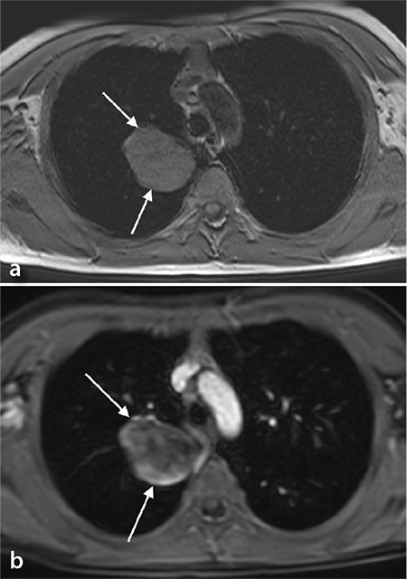
Figure 7.

(a, b) A nine-year-old boy diagnosed with lung adenocarcinoma. (a) The axial T1- weighted image shows a well-circumscribed and isointense tumor in the right upper lobe (arrow). (b) Note the heterogenous enhancement on the T1-weighted post-contrast image (arrow).
