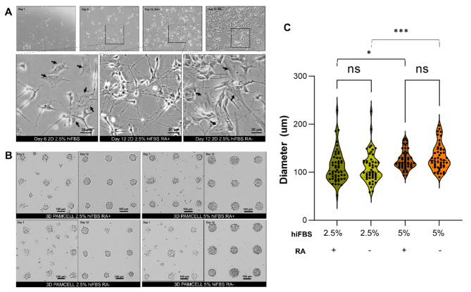
Fig. 1.
Morphology of differentiated SH-SY5Y cells in 2D and 3D PAMCELL cultures with/without RA in maturation media and different serum conditions. (A) Bright-field images of SH-SY5Y in 2D culture on day 1, day 6, and day 12. The arrow (black) indicates the S-type cells, which is flat and stretch on surface. Images were captured using an inverted epifluorescence microscope at 60X magnification in phase contrast. (B) Bright-field images of SH-SY5Y spheroids on day 1 and day 12. (C) Violin plots of the size of spheroids cultured in different serum concentrations. Diameters of 50 random spheroids were measured. Significant differences between different cultured conditions were evaluated. (ns = not significant, *p < 0.05, ***p < 0.001, unpaired Student’s t-test).

