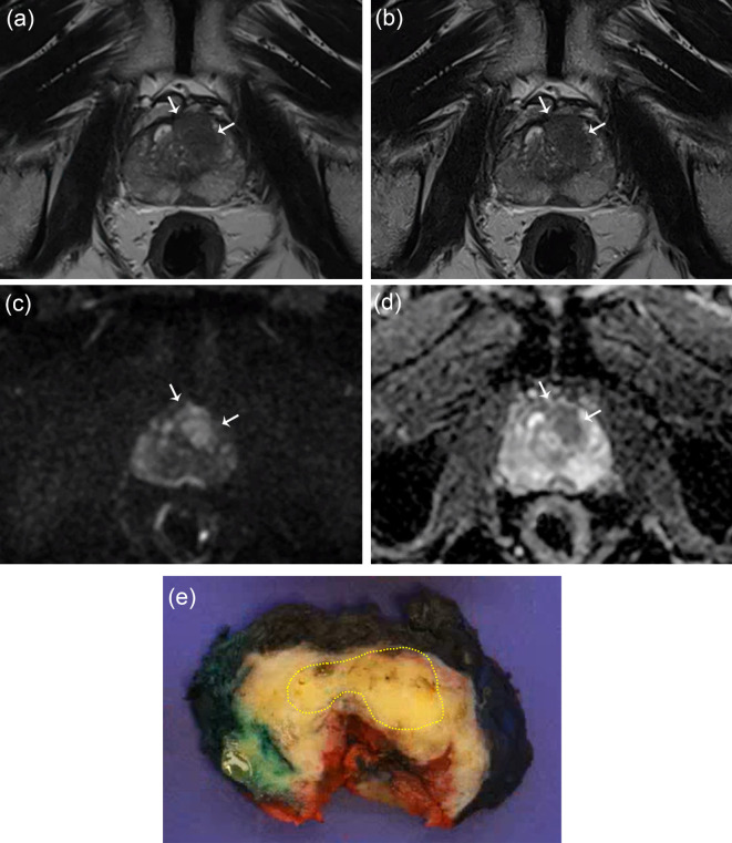
Fig. 2.
A 67-year-old man with a prostate-specific antigen level of 12 ng/mL. An ill-defined lesion with a homogeneously low signal intensity is observed in the left mid-anterior transition zone and mid-anterior peripheral zone on turbo spin echo T2-weighted imaging (T2WI) (a) and deep learning-reconstructed T2WI (b). This lesion exhibits high signal intensity on diffusion-weighted imaging (DWI) with a b value of 1400 s/mm² (c) and shows a low apparent diffusion coefficient (ADC) (d). Both radiologist 1 and radiologist 2 classified the lesion as having a PI-RADS score of 5 on both T2WI sequences and evaluated it as extraprostatic extension (EPE) grade 1. The lesion was histologically confirmed as Gleason grade group 2, and EPE was verified at the anterior aspect of the lesion (e).

