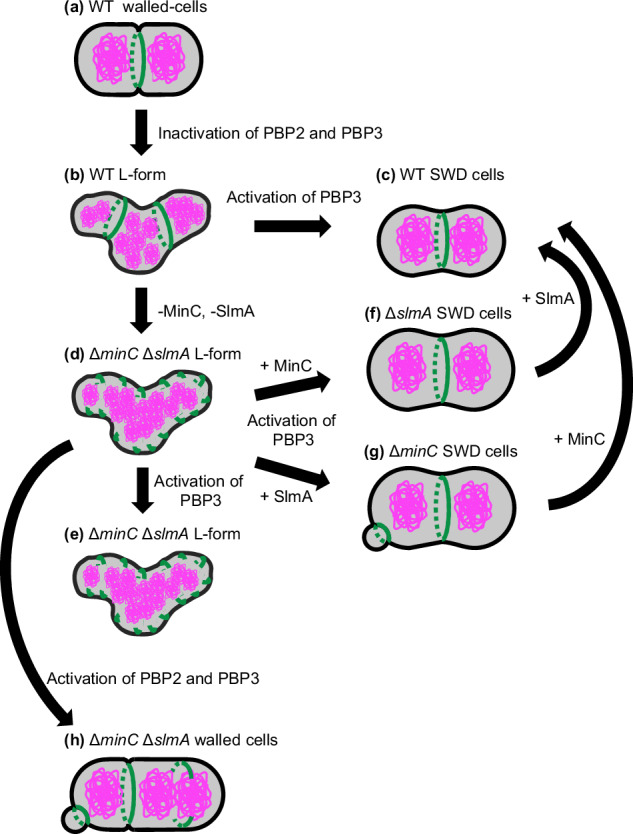
Fig. 6. Model for cell shape control and Z ring formation in SWD cells.

In WT walled-cells, FtsZ (green) formed the Z ring between the segregated nucleoids (magenta) (a). In the WT L-form cells, the Z rings containing all components of the divisome proteins were formed in the nucleoid-free region and could not constrict (b). WT SWD cells were divided into uniform oval cells using Z-ring by the activation of PBP3 (c). In the ∆minC ∆slmA L-form, the short green lines show incomplete FtsZ rings or filaments. ∆minC ∆slmA L-form cells failed to divide in an FtsZ-dependent manner after the activation of PBP3 (d, e). In ∆slmA SWD cells and ∆minC SWD cells, Z ring formation was determined by the Min system and nucleoid occlusion system, respectively. In ∆slmA cells, the Z ring was formed at mid-cell by using only the Min system (f). While in ∆minC cells, the Z ring was formed at cell-pole and mid-cell but not on the nucleoids. Constriction of the Z-ring at the cell poles produces minicells of SWD lacking a nucleus (g). In ∆minC ∆slmA cells, cells were initiated to divide and converted to rod-shaped cells by activation of PBP2 and PBP3 (h).
