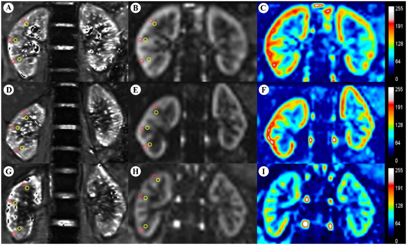
Figure 2.
BOLD-MRI T2* map (A,D,G) and ASL-MRI pCASL map (B,E,H) and false color map (C,F,I) in the coronal plane of the HVs and CKD patients. ROI-based technique, with placement of circled ROIs in the renal cortex (red) and medulla (yellow). (A–C) HV, 42-year-old woman. (D,E) Patient with CKD stage 2, 28-year-old man. (G–I) Patient with CKD stage 4, 57-year-old man. BOLD-MRI: blood oxygen level-dependent magnetic resonance imaging; ASL-MRI: arterial spin labeling magnetic resonance imaging; CKD: chronic kidney disease; HVs: healthy volunteers; ROI: region of interest.

