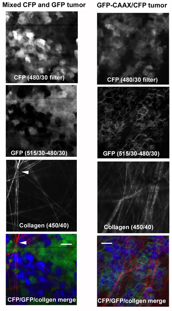
Figure 3.
Simultaneous capture of GFP, CFP and collagen second harmonic fluorescence in a living tissue. The panels on the left-hand side show images of an experimentally generated mammary tumour creating by injecting a mixture of cells either expressing GFP or CFP. The panels on the right-hand side show images of an experimentally generated mammary tumour with cells expressing CFP to mark the entire cytoplasmic volume and GFP-CAAX (membrane targeted). 880 nm laser light was used to excite the all the samples and the fluorescence was captured with indicated filters using non-descanned detection. All images were taken with a 40× objective and a final magnification of 500×. Scale bar = 25 μm.

