
FIGURE 1.
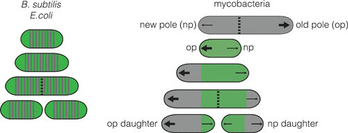
Characteristics of growth and division in B. subtilis and mycobacteria. B. subtilis and E. coli grow by adding new cell wall (gray) along the lateral cell body. Mycobacteria grow only at the polar regions, and do so at unequal amounts depending on the identity of the pole. This is observed by using a cell wall dye (green) to stain the existing cell wall and observe outgrowth of the newly synthesized, unstained cell wall (7). Arrows, polar location of new cell wall synthesis (a large arrow indicates more growth); dotted line, septum; green portion, old cell wall; gray portion, new cell wall.