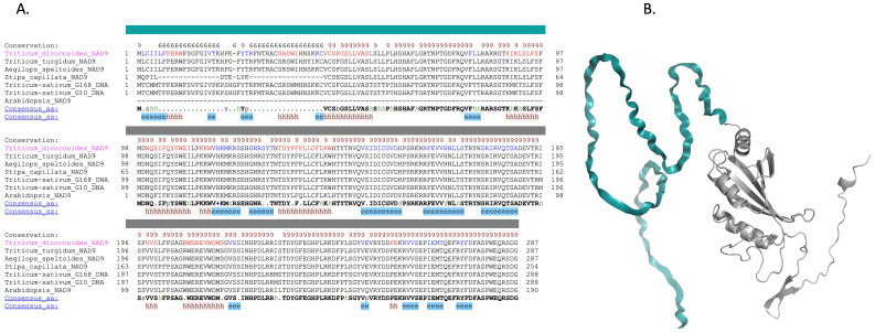
Figure 7.
Comparative analysis and structural modeling of the NAD9 protein across species. (A) Multiple sequence alignment of the NAD9 protein from seven diverse species, highlighting a 98-amino acid (in dark color) region unique to Triticum aestivum. The alignment is annotated to indicate conserved alpha helices (represented by ‘h’) and beta strands (‘e’), with conserved residues highlighted against the consensus sequence at the bottom. Complete conservation across species is denoted by an asterisk above the alignment. (B) Ribbon diagram depicting the NAD9 protein structure in Triticum aestivum, detailing the arrangement of alpha helices and beta strands corresponding to the annotated sequence alignment.

