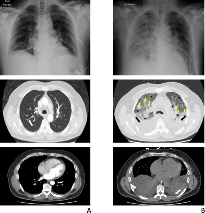
Fig. 1.
Chest X-rays and chest computed tomography (CT) scans. A Initial radiologic studies. Fibro atelectatic tracts compromising the posterior segments of bilateral inferior lobes (white arrow). B. Control radiologic studies (taken after respiratory deterioration). Bilateral basal consolidations involving all segments of the lower lobes (black arrows), associated with posterior pleural effusions (white arrows). Extensive patchy opacities in the periphery of both lung fields (yellow arrows). Ground glass pattern in the remaining lung fields (black asterisks)

