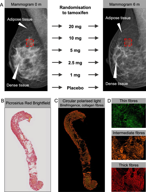
Fig. 1.

Study design. A Mammograms taken before and after randomisation and treatment with tamoxifen at five doses or placebo for 6 months and illustrating the site of biopsy (red square), dense glandular and stromal tissue (white), and non-dense fatty tissue (dark). B, C A biopsy stained with picrosirius red and imaged with B bright-field light microscopy or C circularly polarized light (cPOL) microscopy. D Representative inset images of machine-learning-based segmentation of fibre classes from cPOL images (green = thin fibres, yellow/orange = intermediate fibres, red = thick fibres)
