. 2024 Sep 29;11(3):655–663. doi: 10.5455/javar.2024.k815

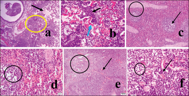
Figure 2. In H&E satin, the lung tissues showed hemorrhages with massive infiltration of reactive cells (10X, a, circle), mainly lymphocytes and neutrophils (40X, b, black arrow), and hemorrhages from the alveolar-capillary (40X, b, blue arrow). The lung’s alveoli were sunk in exudate and appeared light pink (10X, a, arrow). There was lymphoid depletion (10X, c, arrow and 40X, d, circle) and enlargement of trabeculae (10X, c, circle) in the infected spleen. Lymphoid depletion was also observed in mesenteric lymph nodes (4X, e, circle and 40X, circle). There were congestion and hemorrhages in mesenteric lymph nodes (40X, b, arrow).

Figure 2.