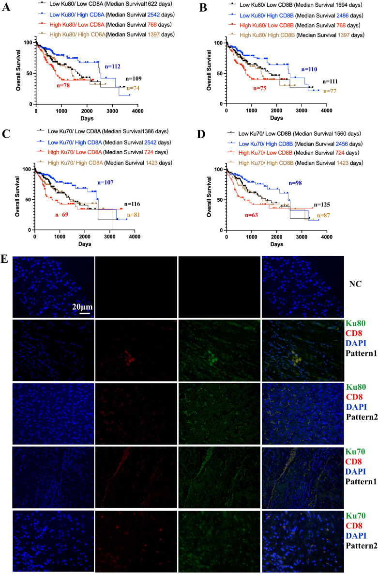
Figure 4.
Combined analysis of Ku70, Ku80, CD8+ T cells in TCGA database and their tissue expression pattern validated by co-localized immunofluorescence staining. (A). Combined analysis showed the expression of Ku80 and CD8A and the survival outcomes in HCC patients (log-rank p=0.0014). High Ku80/low CD8A vs low Ku80/high CD8A showed HR=2.64 (95% CI 1.532 to 4.549, log-rank p=0.0003). (B). Combined analysis showed the expression of Ku80 and CD8B and the survival outcomes in HCC patients (log-rank p=0.0023). High Ku80/low CD8B vs low Ku80/high CD8B showed HR=2.504 (95% CI 1.462 to 4.289, log-rank p=0.0002). (C). Combined analysis showed the expression of Ku70 and CD8A and the survival outcomes in HCC patients (log-rank p=0.0023). High Ku70/low CD8A vs low Ku70/high CD8A showed HR=2.713 (95% CI 1.566 to 4.699, log-rank p<0.0001). (D). Combined analysis showed the expression of Ku70 and CD8A and the survival outcomes in HCC patients (log-rank p=0.0019). High Ku70/low CD8B vs low Ku70/high CD8B showed HR=2.763 (95% CI 1.566 to 4.877, log-rank p=0.0001). (E). immunofluorescence co-localization showed the different expression patterns Ku70/CD8+ CTL and Ku80/CTL. Ku80/Ku70/CD8+CTLs/DAPI.

