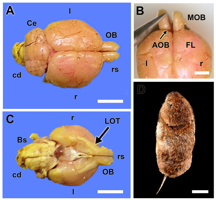
Figure 1.
Macroscopic anatomy of the olfactory bulb of the fossorial water vole. (A) Dorsal view of the brain showing the relative size of the olfactory bulbs (OB). (B) Dorsal view of the rostral part of the telencephalon showing the location area of the AOB. (C) Ventral view of the brain showing the remarkable size of the lateral olfactory tract (LOT). (D) Dorsal view of one of the analyzed specimens of fossorial water vole. Bs: Brainstem; cd: Caudal; Ce: Cerebellum; FL: frontal lobe; l: left; MOB: main olfactory bulb; r: right; rs: Rostral. Scale bars: (D) = 2 cm, (A,C) = 1 cm, (B) = 0.5 cm.

