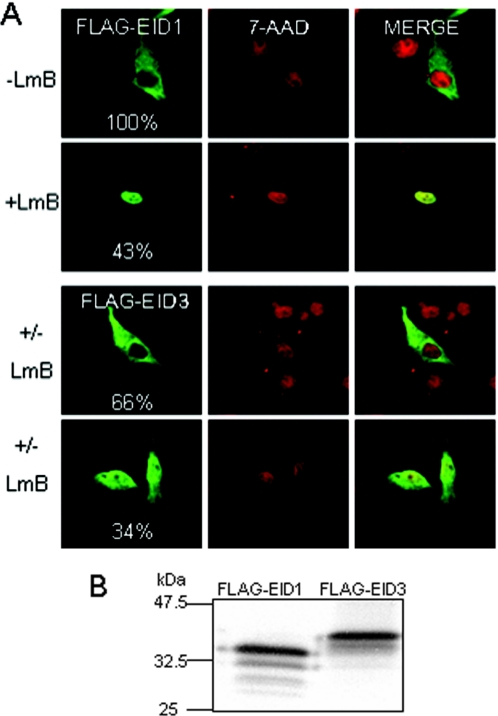
Figure 2.
Sub-cellular distribution and protein analysis of EID3. (A) Intracellular localization of EID1 and EID3. FLAG-tagged EID1 and EID3 were expressed in COS-7 cells and analysed by indirect immunofluorescence using FLAG antibody (green) in the absence or presence of LMB (5 nM for 5 h). Nuclei were stained with 7-aminoactinomycin D (7-AAD) (red). More than 50 cells were studied and the experiment was independently reproduced at least three times. (B) In vitro transcribed and translated [35S]methionine-labelled EID1 and EID3 are shown.

