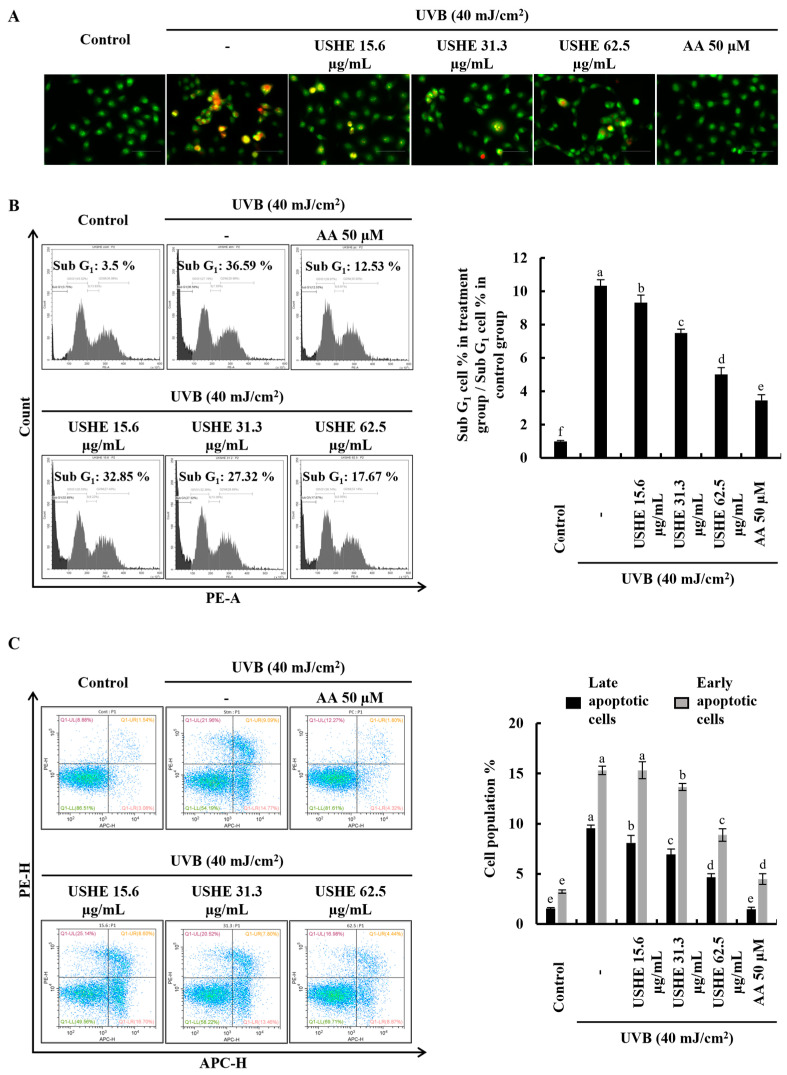
Figure 3.
Effect of USHE on the apoptotic cell population in UVB-exposed HaCaT keratinocytes. (A) Nuclear morphology analysis using EB and AO double staining (Objective magnification of the instrument: 40×), (B) sub G1 apoptotic population analysis by flow cytometry using PI, and (C) early apoptosis investigation using flow cytometry Annexin V analysis. Values are expressed as the mean ± SE from triplicate experiments. Bars with different letters within the same graph represent significant differences (p < 0.05).

