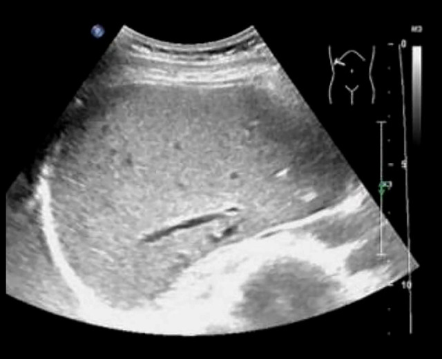
Figure 3.

Liver ultrasound of the patient, performed 3 months after surgery. Three months after surgery, the patient came to our outpatient department for re-examination by abdominal ultrasound. The results indicated that the liver surface was smooth, the liver margin was sharp, and no obvious lesions were found where the hematoma had been detected by computed tomography scan during hospitalization, suggesting that the patient’s subcapsular liver hematoma and gas accumulation had been completely absorbed.
