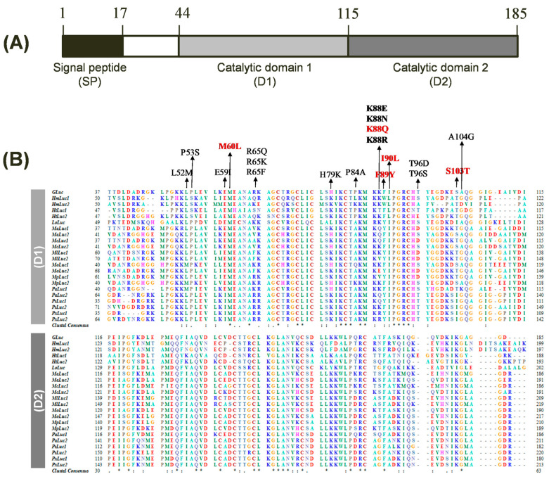
Figure 1.
Structure and sequence alignment of Gaussia luciferase (GLuc). (A) Domain architecture of GLuc encompassing a signal peptide (SP) and two repeated catalytic domains (D1 and D2). The protein comprises 185 amino acids. (B) Multiple sequence alignment of GLuc and 21 copepod luciferase homologs, focusing on the D1 (top) and D2 (bottom). Amino acid residues are color-coded based on their physicochemical properties. Identical residues across all sequences are indicated by asterisks (*), while conserved substitutions are represented by a colon (:) and period (.). Gaps introduced for optimal alignment are shown as dashes (–). Amino acid positions are numbered on the left and right. The amino acids targeted for mutagenesis are indicated by arrows, with GLuc5 mutation sites highlighted in red.

