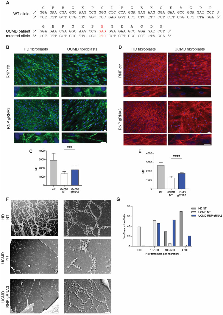
Figure 4.
Secretion of collagen VI in fibroblasts from healthy donor (HD) and UCMD patient after treatment with RNP. (A) Nucleotide and amino acid sequence of exon 9 in UCMD patient carrying c.824_838del variant. In red, the insertion of a Glu residue is shown. (B) Immunofluorescence analysis with antibody specific for collagen VI (MAB1944) of fibroblasts from HD or UCMD patient, treated with control RNP (RNP ctr) or RNP-gRNA3. The experiment was performed in quadruplicate. A representative experiment is shown. Enlarged image insets show the structure of collagen VI microfibrils. Scale bars indicate 20 µm magnification. (C) Mean intensity of collagen VI quantified in fibroblasts treated with RNP-gRNA3. Data represent mean ± SD from analysis of 10 individual field images acquired at 100× original magnification under fluorescence microscopy. *** p < 0.001). (D) Immunofluorescence analysis with collagen VI-specific antibody (polyclonal Fitzgerald) of primary fibroblasts from HD or UCMD patient treated with control RNP (RNP ctr) or RNP-gRNA3. The polyclonal Fitzgerald anti-collagen VI antibody was used to label α1, α2 and α3 chains of collagen VI. A representative experiment is shown. Enlarged image insets show the structure of collagen VI microfibrils. Scale bars indicate 20 µm magnification. (E) Mean intensity of collagen VI quantified in fibroblasts treated with RNP-gRNA3. Data represent mean ± SD from analysis of 10 individual field images acquired at 100× original magnification under fluorescence microscopy. **** p < 0.0001). (F) Ultrastructural analysis by rotary-shadowing electron microscopy of collagen VI microfibrillar network in untreated (NT) HD fibroblasts, UCMD fibroblasts treated with RNP-gRNA3 or not (UCMD NT). (G) Percentage of microfibrils composed by increasing amounts of tetramers in UCMD fibroblasts treated with RNP-gRNA3 or not (UCMD NT) and in control fibroblasts (HD NT).

