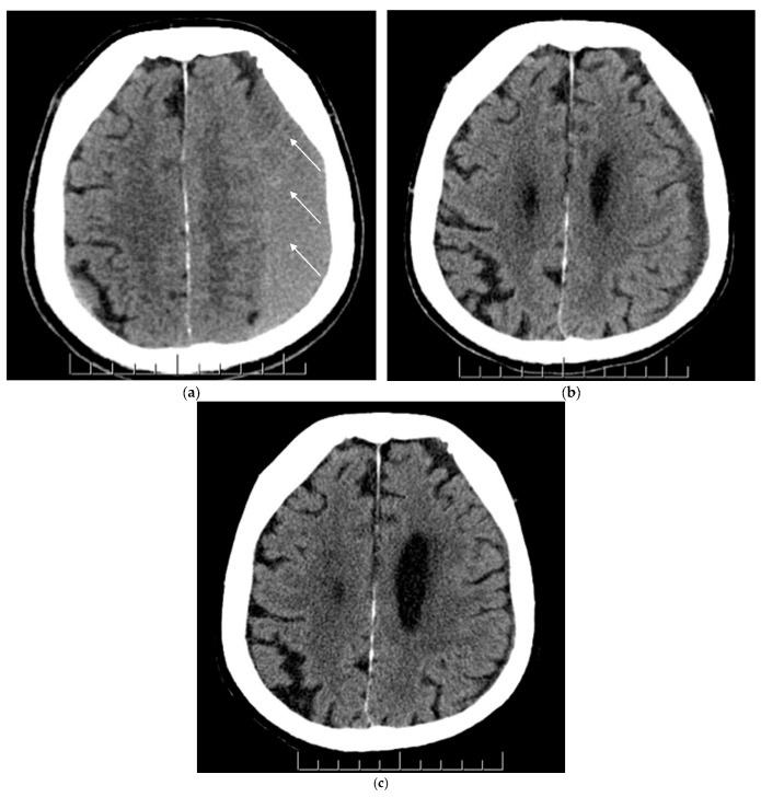
Figure 1.
(a–c) Axial view of non-contrast CT head scans before and after MMAE (middle meningeal artery embolization) for the same patient. Figure parts a, b and c represent chronic subdural hematoma (cSDH) pre-MMAE (baseline), at 1–3 months and 3–6 months post-MMAE, respectively. (a) A non-mature mixed cSDH was noticed at baseline on the left cerebral hemisphere with sulcal effacement, obliteration of the left lateral ventricle and slight midline shift to the right. The white arrows demonstrate the mixed cSDH nature; (b) Improvement of the cSDH and mass effect was noticed in 6-week CT scan post-MMAE; (c) A near-complete resolution of cSDH and mass effect was noticed in 5-month CT scan.

