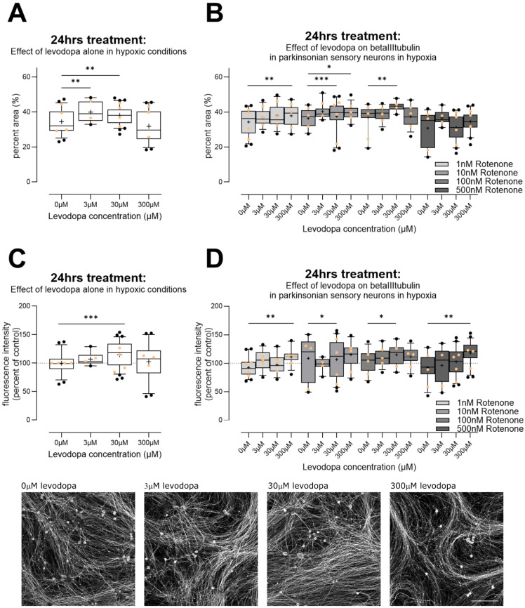
Figure 5.
Effect of levodopa on beta III tubulin in primary sensory neurons (DRGs) cultured in hypoxia. (A) Alone, 3µM and 30 µM levodopa increased percent area immunoreactive for beta III tubulin. (B) In the context of parkinsonian sensory neurons (treated with rotenone), levodopa tended to increase percent area positive for beta III tubulin at 30 µM and 300 µM. (C) We examined fluorescence intensity for beta III tubulin, at the level of individual neurites in cells. Cells treated with levodopa only again showed increased fluorescence at 30 µM. (D) In the context of parkinsonian sensory neurons (treated with rotenone), 30 µM and 300 µM levodopa tended to increase fluorescence for beta III tubulin. Data are technical replicates shown as box plots with whiskers depicting 5–95% percentiles, black circles depicting remaining data points, lines depicting medians and “+” symbols depicting means. Light orange circles depict experimental means. * p < 0.05, ** p < 0.01, *** p < 0.001 post hoc tests, following ANOVA, as discussed in text. Photomicrographs show example original images of cells treated for 24 h in hypoxia with 0 µM, 3 µM, 30 µM, 300 µM levodopa, only, then stained for beta III tubulin. Photomicrographs are not modified. Scale bar bottom right = 200 µm, for all images.

