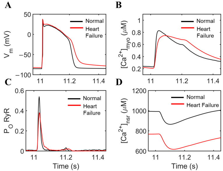
Figure 1.
Comparisons of normal cardiac cell conditions vs. heart failure. To ensure steady state conditions, 20 s simulations at 1 Hz were performed. (A) APD prolongation with longer plateau phase due to influx of Ca2+. Early afterdepolarizations (EADs) were also observed through the oscillations of the membrane potential. (B) Ca2+ concentration displays an incomplete extrusion from the myoplasm. (C) RyR2 open probability is decreased during heart failure, and small openings were observed during EAD occurrence. (D) SR Ca2+ also displays incomplete recovery, mainly due to decreased SERCA activity and increased NCX expression.

