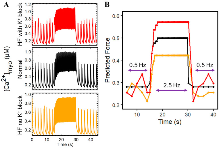
Figure 3.
Dynamic slow–rapid–slow pacing (0.5–2.5–0.5 Hz) [Ca2+]myo concentrations between normal, HF with K+ block (i.e., Ito, IK1, IKr, and IKs), and HF without K+ block. To ensure steady state conditions, 45 s simulations were performed. (A) Erratic Ca2+ transient peaks were observed during 0.5 Hz pacing in failing hearts. HF with reduced K+ currents (red) slightly elevated the [Ca2+]myo amplitude during high pacing (2.5 Hz) as compared to normal (black) and HF without K+ reductions (yellow). (B) Predicted force using the same color schemes. HF with reduced K+ currents (red) exhibit higher predicted force using peak systolic [Ca2+]myo concentrations in rapid pacing.

