Figure 4.
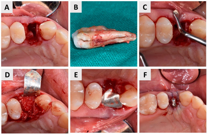
Magnesium membrane shield technique for alveolar ridge preservation (ARP) in complete buccal bone loss (buccal dehiscence); (A) Occlusal view of the extraction socket. After administration of local anesthesia, the tooth was extracted as atraumatically as possible. The socket was carefully debrided using a surgical curette, with a finger placed buccally to protect the gingiva, which lacked an underlying bone base; (B) The extracted tooth (24, FDI notation) showing a visible vertical root fracture; (C) Assessment of the buccal mucosa with an instrument, confirming the absence of the buccal bone wall; (D) A gentle elevation of the soft tissue was performed on both the buccal and palatal sides to facilitate the insertion of the magnesium membrane (NOVAMag® membrane, botiss biomaterials GmbH, Berlin, Germany). After the socket was filled with a bovine-derived xenograft (cerabone®, botiss biomaterials GmbH, Zossen, Germany), the membrane was positioned between the bone graft material and the soft tissue following the contours of the pre-existing buccal wall; (E) The magnesium membrane was then carefully shaped to curve over the grafted area and positioned beneath the palatal gingiva, adjacent to the palatal bone wall; (F) Finally, the membrane was stabilized and secured in place with 6-0 sutures (Luxylene, Weiswampach Luxembourg), allowing healing via secondary intention without primary wound closure.

